Back to Top
Augusta University LogoShield

Information for:

  • Current Students
  • Alumni
  • Community
  • Faculty & Staff
  • 伟德betvicror
  • Patients

Resources

  • 伟德APP
  • Calendar
  • 伟德1946英国
  • D2L LMS
  • Email
  • 伟德1946客户端下载
  • MyAugusta
  • News
  • 伟德1946娱乐城
  • Portals
  • Pounce
  • 伟德1946网页版
  • Admissions
  • 伟德1946亚洲娱乐城
  • About
  • Student Life
  • Athletics
  • Research
  • Health
  •   Giving
Medical College of GeorgiaCell Imaging Core | Cellular Biology & Anatomy
Cell Imaging Core | Cellular Biology & Anatomy
  • About
  • Fees
  • Contact
  • Rules
  • Instrumentation
  • Calendars
  • Useful Links
  • FAQ
  •  

  • Augusta University
  • Colleges & Schools
  • Medical College of Georgia
  • Cellular Biology & Anatomy
  • Cell Imaging Core | Cellular Biology & Anatomy

Cell Imaging Core

The Cell & Tissue Imaging Core Laboratory is committed to excellence in research and education. We provide expert technical assistance and instrumentation in support of investigators.

   The Core educates graduate students in the basic principles of imaging.  


Publication Requirements: To cite or acknowledge the use of our core: Augusta University Medical College of Georgia Cell Imaging Core Facility, RRID:SCR_026799
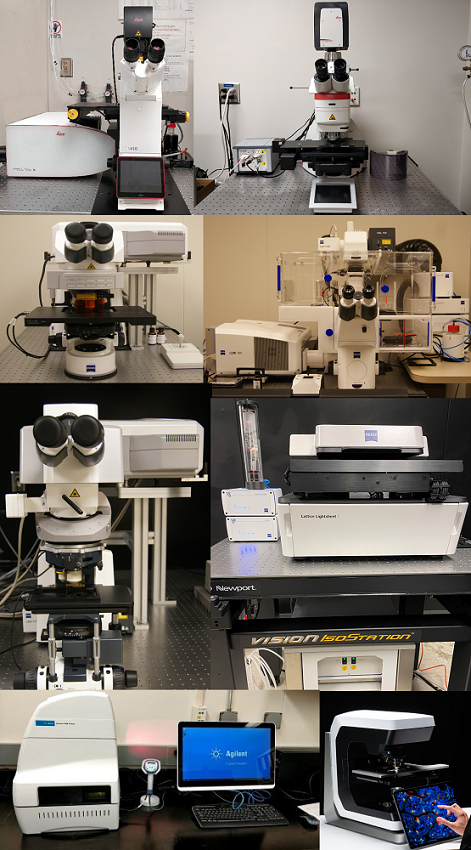
Fees
Learn More

Instrumentation

The Cell Imaging Core Laboratory has a variety of microscopes available for use.

link
cell core image
Learn More

Rules

All new users are required to be trained via the Imaging Core staff prior to using an instrument unless they can demonstrate instrument competency.

Link
Fees
Learn More

Fees

There is currently an $21.30 per hour instrument usage fee for most instruments. Utilization of these instruments is reserved for Augusta University researchers only.

Link
FAQ
Learn More

FAQ

Have questions? Check here first!

Link
Calendars
Learn More

Calendars

Steps on how to schedule & kiosk instructions.

Link
Contact us image
Learn More

Contact

Contact us for any questions!

Link

The Cell and Tissue Imaging Core Laboratory has the following research capabilities:

Located in CB-2309:generic cells under a microscope

  • High-performance Digital Slide Scanner for Fluorescence, Brightfield and Polarization: Zeiss Axioscan 7 Digital slide scanner
  • Confocal microscopy and 3D visualization: Leica Stellaris 5, Zeiss LSM780 upright and inverted confocals
  • Multiphoton microscopy: Zeiss LSM780 upright multi photon, Nikon AXR upright multiphoton
  • Super-resolution microscopy: Nikon STORM, Zeiss LSM780 inverted with Airyscan, Leica Stellaris 5 with Lightning
  • Live imaging of cells and tissues: Zeiss Lattice Lightsheet, Leica Stellaris 5, Zeiss LSM780 inverted
  • Laser capture microdissection: Leica LMD6
  • Image analysis using Imaris 10.1 software (Oxford instruments)
  • Measurement of oxygen consumption rate and extracellular acidification rate of live cells on a 96 well plate format: Agilent Seahorse Xfe96 Analyzer

Located within the animal facility in the Georgia Cancer Center (CN-5113):

  • Live animal microscopy: Nikon AXr-Multiphoton Intravital microscope

The core is directed by Dr. Graydon Gonsalvez. Daily operations & maintenance are the responsibility of Dr. Rachel Cui.

photo of Dr. Graydon Gonsalvez

Dr. Graydon Gonsalvez

  • Director, CIC
  • Associate Professor, CBA

706-721-2551

imagingcore@augusta.edu

photo of Dr. Rachel Cui

Dr. Rachel Cui

  • Research Manager, CBA

706-721-1876

imagingcore@augusta.edu

Great Doctors, Great Medicine Since 1828. 

University Shield

Augusta University

1120 15th Street, Augusta, GA 30912

  •   Campus Maps
  •   Campus Contacts
  • 伟德APP
  • Degrees & Programs
  • Employment
  • Accessibility
  • Accreditation
  • Campus Safety
  • Compliance Hotline
  • Human Trafficking Notice
  • Privacy Notices
  • Title IX / Sexual Misconduct
伟德1946手机版 Give Now

© 2025 Augusta University

Facebook Twitter LinkedIn Youtube Instagram
©
Baidu
map